


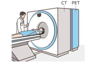
Positron emission tomography is a computed tomography technique that uses positron detection. Also known as
positron emission tomography, or PET. When radioactive substances and glucose-containing drugs enter the body,
glucose-containing drugs accumulate in cancer cells, which have an active metabolism. PET scanners specifically
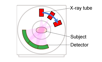
look at how the body functions by detecting positrons. Unlike CT, which examines part of the body, the whole body
can be examined at once. Because it can look at metabolic levels in the central nervous system, it is also used to
diagnose epilepsy and Alzheimer's disease.
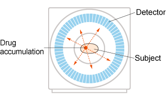
In recent years, it has been used in cancer screening by detecting elevated levels of glucose metabolism in tumor
tissue. Because it can screen the entire body, it is also used to detect cancer metastases and unexpected cancers.
The method used for cancer screening is called FDG-PET, in which a radioactive substance with a structure similar
to sugar is injected into the body, which cancer cells use to metabolize, and its movement is observed. It is
characterized by easy detection of active cancers and effective treatment. It also has the advantage of less radiation
exposure to patients than CT, which uses a wider radiation range.
PET scanners use a special camera that makes use of scintillators, a substance that converts radiation into light. The
camera captures the small amount of light produced by the scintillator and amplifies it to produce an image that is
easy to view and understand.
There are various ways to convert the light emitted by the scintillator into electricity, such as using an image
intensifier (II), photomultiplier tube (PMT) or APD (avalanche photodiode) and MCP (microchannel plate). These
detectors require a high voltage of 100 to 5,000 V.
Wisman offers a variety of high voltage power supplies for PET scanners: MM; MMC; MG; MMA; MCE;